
Recently, we had one of our feline patients come in to get a limp checked out. Art, a middle aged Domestic Shorthair had some arthritis in his knee causing his limp (arthritis in cats is a separate blog post all together, and we will be addressing it in a few weeks) but when Dr. T checked his mouth (as part of her routine exam) she found something much more worrisome. Art had large holes in his teeth… and in some cases, entire teeth were just … missing!
Dental disease is a VERY general term and is one of the most common (if not THE most common) ailment of pet dogs and cats. Most pet owners will have heard their vet talk about gingivitis, tartar build up, maybe even gum recession and periodontal disease when examining their pet’s teeth and gums – these are the most common dental issues we see in pets. One of the more serious and complex diseases we see when it comes to your pet’s mouth is Tooth Resorption.
What is Tooth Resorption?
Most commonly seen in cats (but can happen in dogs too) this is a progressive disease where the tooth is actually eaten away or dissolved by the pet’s body – weird right? It can happen in people too, but is much more common in our feline friends. This can become a very painful condition for your pet. Their teeth and gums become sore and it hurts to chew. Animals have an incredible ability to hide their discomfort though (evolutionary advantage to not show your predators that you are ill or weak) and so sometimes pet owners won’t have any outward signs that this is going on.
It could happen on the crown of the tooth (the visible part that sits above the gum line) and we can actually see the tooth being eaten away, like the picture below.
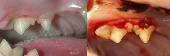
When we see lesions like these, we know we have some work to do.
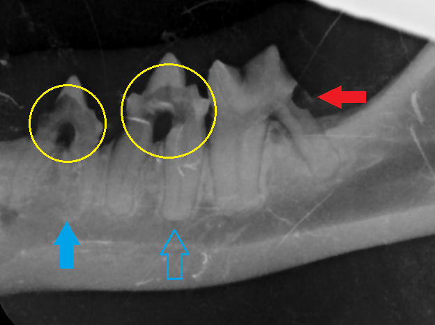
The tricky part about tooth resorption is that sometimes the crowns of the teeth look fairly normal and it is the root lying under the gum that is affected. The only way we can diagnose this type of resorption is with dental radiographs. This is why it so important to do full mouth radiographs on any animal showing other signs of tooth resorption. Look at the picture below – a radiograph of a patient we had in to the clinic a few weeks ago, Art.
We’ll try not to get too in depth into dental anatomy and physiology but we’ll go over the basics.
Red arrow- An area of resorption that we couldn’t see during Art’s physical exam – the hole in the tooth is sitting just at the gum line on a back molar, facing the back of the mouth – a very difficult place to assess even in the most cooperative of patients.
Yellow circles – See how the tips of the teeth are brighter white than the centres? That’s because the tooth is thinner in the darker grey area. This is erosion on the crowns of the teeth – these were visible in his exam, and the main reason for recommending dentistry for further examination.
Blue arrows – Compare the roots of the teeth between the solid and outlined arrows. The outlined arrow shows a healthy root – you can follow the outline and can clearly see tooth root vs. surrounding jaw bone. The solid arrow shows resorption of the roots where that clear definition is lost.
In Art’s case, all 3 of these teeth needed surgical extraction. Luckily for Art, his mom ok’d the dental work and he is now home, pain free, and recovering well from his surgery.
And here’s the most frustrating part for us – for the most part, we can’t find a cause for tooth resorption. Some cases start where there has been obvious damage to the tooth and the living part of the tooth – the pulp – actually dies, but for the most part, we don’t know why it happens – which means we can’t know who it is going to affect or prevent it from happening.
This is just one more thing that can be checked and diagnosed early with regular check ups with your pets and a good reason to take your indoor cats in for their annual exam even though they may seem healthy.